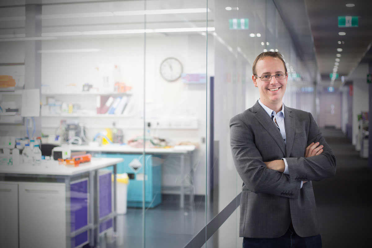
Introducing Associate Professor Andrew Bivard
In a world of health care where terms like artificial intelligence and machine learning are bandied about, the Australian Stroke Alliance is cutting out the middleman and shooting straight for a cloud when it comes to real time, personalised care of patients in remote locations.
Here, we meet a member of our team, Andrew Bivard, who is based at the Melbourne Brain Centre at the Royal Melbourne Hospital and at The University of Melbourne. He’s pushing the limits of stroke perfusion imaging to enhance diagnosis and to fast track treatment.
Andrew is one half of the self-described Odd Couple with neurologist Mark Parsons and, together, they are developing novel telehealth technology to connect doctors to their patients, no matter where they may be, on the mobile stroke unit or in a regional centre.
Ok Andrew Bivard. Elevator pitch, please. Tell us about your research and the way you are improving support for paramedics, nurses and GPs in rural and remote locations. You have 30 seconds and your time starts now.
Over the last five years there has been an explosion of evidence proving that careful patient assessments – supported by brain imaging – guides clinicians in the best treatment pathway for their patients.
I did my PhD in the most versatile of these imaging methods, perfusion imaging, validating it as a way to measure areas of dead brain due to stroke, from areas that can be salvaged with effective therapy.
Differentiating these sections of brain tissue is the cornerstone for extending the treatment time window for thrombectomy to 24 hours, and thrombolysis to nine hours. Our team leads the world in extending these windows for treatment.
Perfusion scanners are expensive and require expertise to run. As a result, imaging is usually performed at regional centres but sometimes, this requires a petition to government to buy a scanner and then for staff to be trained (which they always love as it is a cool new technique for them).
Once this hardware is in, we need to ensure that stroke specialists can access the information it generates and that the information is accurate.
The problem, though, is that stroke physicians are rarely based in rural settings. As well, doctors tell us that their diagnosis and treatment of a stroke patient requires more than just images of the patient’s brain. Ideally, they need to actually see a stroke patient when making a diagnosis.
They also want access to clinical note taking, record keeping and even to be told when a potential stroke patient is on the way to the hospital. To overcome the tyranny of distance, we are building a fully independent, world-first telestroke app.
And the good news is that we have already trialled a version on the Melbourne-based mobile stroke ambulance. Over the last six months, we’ve learnt a lot about what does and does not work. Overall the platform has performed perfectly to expectations.
To be honest, I was just happy that the ‘on’ button worked at day one! We are transmitting crucial clinical data and brain images, as well as audio-visuals of the patient from the vehicle to stroke specialists.
We are moving to an even better version which will be a fully independent and is set to be deployed. Once it is up, running and stable, the fun can begin and we can tack-on great innovations that mine the data that we generate using machine learning.
How can people find out more?
Well, fortunately, Nature Reviews Neurology has released a publication of ours which is a manifesto on how information can be used in the acute stroke pathway, and where different types of information processing such as machine learning can fit in.
The skull is a very effective bony helmet, protecting the brain. Clearly, CT imaging is an essential tool when a medical team is trying to make a diagnosis in the outback. What will be the role of imaging in, say, 10 years’ time? Will it still be needed, or will other technologies and tools replace it? Will AI-based decision-makers depose the expert clinician?
Imaging will always be needed. It is important to confirm and estimate the severity of an illness for guiding treatment and predicting the outcome from treatment, as well as measuring benefit.
History has shown that a clinical assessment alone is a very poor substitute for imaging, a bad migraine has overlapping symptoms of a minor stroke. That said, there could always be some blue-sky technology.
There are microwave helmets being tested now which may be able to replace the humble CT scan, although the helmet will not be able to replace a perfusion scan.
You have a real interest in it and psychology. How did you find your way into neuroscience and who inspired you?
Well neuroscience is just the science part of psychology. I’m sure that might cause some sparks! I actually started uni doing IT, but just could not connect with the content and the teaching method really did not gel with me.
Then I did what every lost uni soul does and swapped to Psychology and really enjoyed the more scientific parts which took me into Neuroscience. I really didn’t start to do well at uni until I took on Neuroscience subjects.
Interested to read more?
Our national, digital telestroke network.
Meet telestroke neurologist, Chris Bladin
Sign up for our newsletter to keep in-touch.